
These last couple of weeks have been really busy – so much to do before the OHBM meeting in Rome! Trying to finish manuscripts, working on scientific posters with colleagues, as well as preparing a couple of talks for Rome – one for an OHBM Symposium I have co-organized & another for a Symposium at the Sapienza Università di Roma.
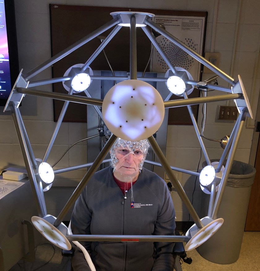
The other thing we have been trying to do is to finish up our study of ‘living phantoms’ – my colleague & I have already made recordings of each other’s brain electrical activity and brain blood flow on the other side of the Atlantic, in my lab & in the 3 T MRI-scanner in our Imaging Research Facility at Indiana University [IU]. So now we are doing the same thing on this side of the Atlantic. The image below shows me wearing our 256-sensor cap [to measure electrical brain activity] at IU. The contraption I am sitting in [image below] is a photogrammetry system in my lab – a device that has 11 cameras that capture a picture of my head & where the sensors are located on the head.
This allows a 3D map of the head surface with sensor positions to be made, which can then be subsequently merged with an anatomical MRI scan of my whole head, which ultimately looks something like this [image below], which was taken a few years earlier.
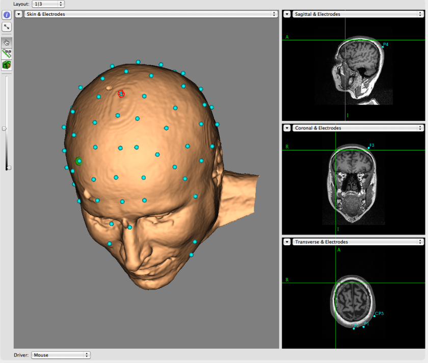
Perhaps you can see that characteristic nose of mine in that hairless & colorless image. If you look carefully you can probably recognize my facial features – if you already know me well. This, of course, is a real ethical issue for subject privacy in labs all over the world. In functional MRI studies it is usual to ‘strip’ away the tissues of the head & face to leave only the brain – which of course is hard to identify. In some of our brain electrical activity [electroencephalography or EEG] studies this is not possible. We need to use the surface of the head/face & also consider how well the different tissues of the head conduct electricity [i.e. the spontaneous brain activity that our brain emits 24/7] for certain types of very specialized data analyses. [This is not the case in all EEG studies – many studies use only the EEG traces & 3D maps of the head are not needed.]
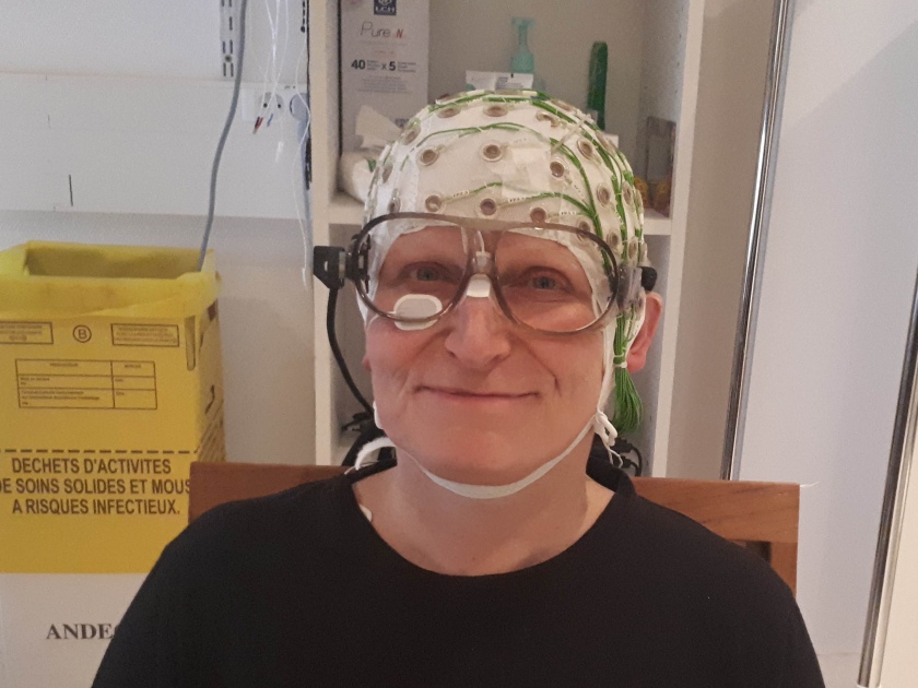
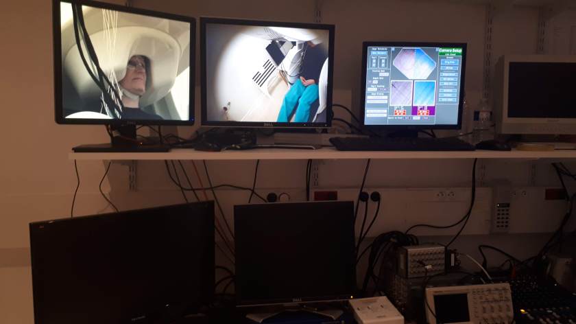
One can also measure the tiny magnetic fields that the brain emits using a method known as magnetoencephalography [MEG for short]. If one wants to really go over the top, one can do both MEG & EEG at the same time. This is what I did this week in CENIR at the ICM. First, the EEG sensors were put on my head, with extra leads to also measure eye movements & cardiac activity. We also need to make a map of the EEG sensors on my head – but here a slightly different method was used. A radio frequency transmitter at the back of the chair I am sitting on [that you cannot see] puts out a signal whose strength varies as a function of 3D distance from it [a polhemus system] . The experimenter uses a wand-like device [a radio frequency receiver] to touch the central location of each EEG sensor & measure the signal strength [& therefore the sensor’s position on the head]. The ‘Biggles’ googles I am wearing also have this position sensing in them, so that if I move my head even slightly the measurement system will compensate for that…
Once we had the sensors localized in 3D space, off came the goggles & it was time to ‘gel’ ’em up i.e. put in some conductive ‘goop’ to provide a good contact between my scalp & each sensor. This is always the fun part & takes quite a bit of time – the goop is usually cold & the experimenters have exfoliate the scalp as they go – yes indeed, one has a spa treatment for the face & head for this experiment… After a lot of checking that the contact between sensors & scalp is good, it is time to go into the shielded chamber, where the recording will take place. There I am fitted into the MEG helmet – a rigid device with sensors embedded in liquid helium. The thing weighs a lot & sits in a gantry that is adjustable. You have to make sure that your head is on contact with the MEG helmet [completely the opposite of being in an MRI scanner where you cannot touch the headcoil or the bore of the magnet].
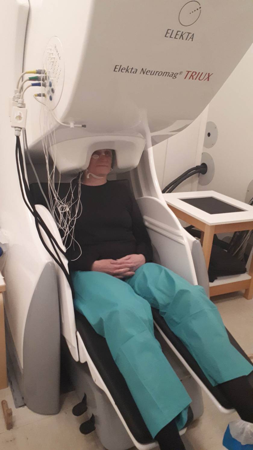
Before we started all of this & changed some of my clothing [PJ pants], took off all of my jewelry & watch etc. We had done a quick check to see that I did not have any residual magnetization in my body [this includes clothing such as metal clips on bras etc & also sometimes dental fillings, for example]. If there is something magnetic on the person’s body – such as clothing etc. one would have to change completely into the PJ set that is supplied. Similarly, shoes also come off & little booties are issued. For dental fillings that are a problem the head can be ‘degaussed’ using a wand-like device – makes me think of ‘aura cleansing’ when I see it done. 🙂 This time I did not need to be degaussed even though I was expecting to have to do so because I had an MRI scan at IU as part of this study & made sure that it was done at least 6 weeks before having this MEG study…
The shielded room is a minimalist place to be sure – nice clean white interior with minimal clutter. There is no electrical equipment is inside the room – the idea is to keep all magnetic fields out – including that from our own planet. This is because the magnetic fields we measure from the brain are really tiny & are indeed many, many orders of magnitude smaller than the Earth’s magnetic field… The shielded room is also soundproof, once the door is sealed, so all communication with experimenters occurs via a MEG-compatible intercom system. In the control room, the experimenters can monitor what I am up to in there at all times – cameras monitor me, as well as all the sensors whose activity is displayed on monitors.
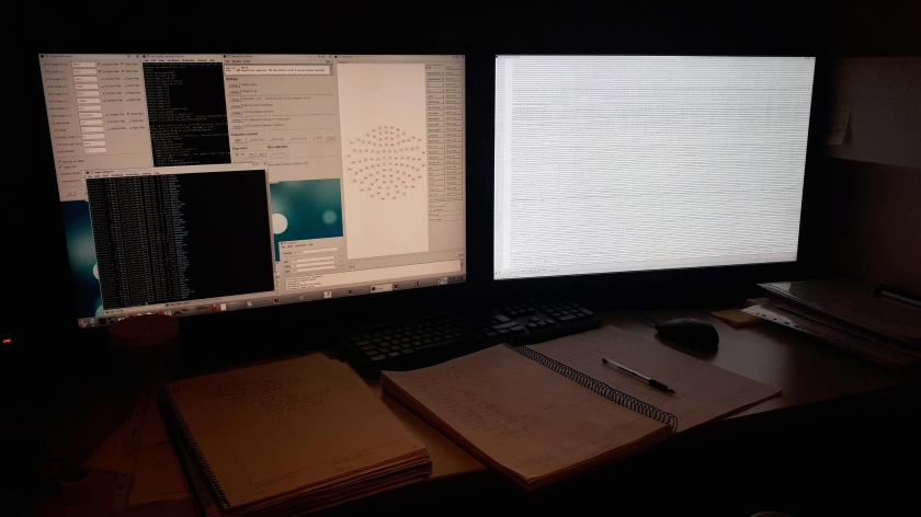
We were doing a ‘resting state’ study on me & also my colleague across 2 different continents – so we are being studied on either side of the Atlantic with multiple assessment modalities. We will look to see how we can integrate these datasets & also look for how consistent the profiles of activity are across the two measurements. Our plan is to share the data with colleagues in an open science framework, but we will have to do some gymnastics with data formatting first, so that it is in a new and desirable format [BIDS] that will allow more people to interact with it.
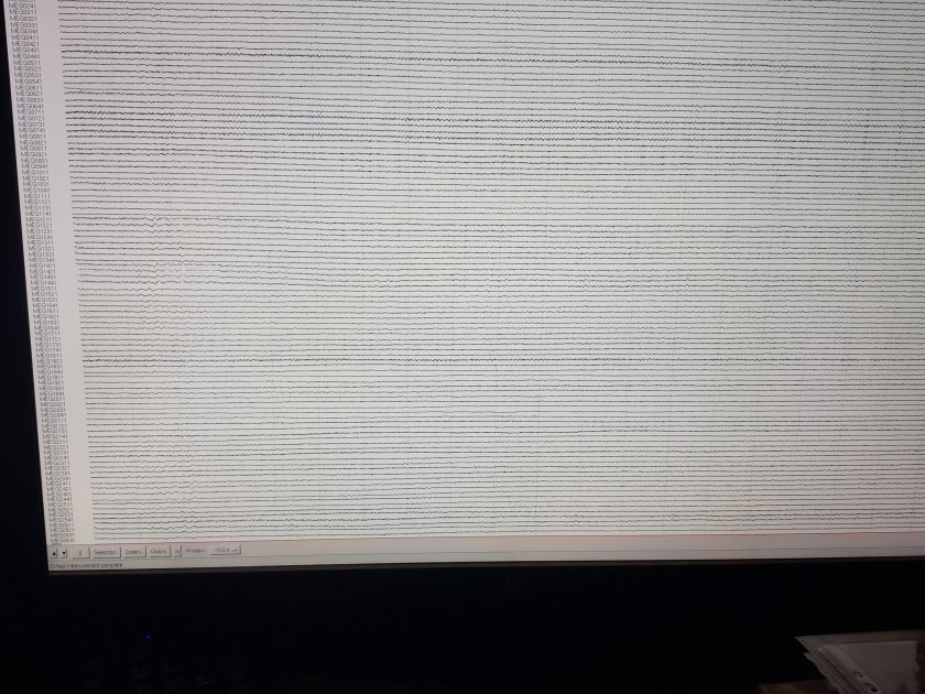
One of the tough things about doing a resting state study is ‘staying on task’ i.e. keeping one’s eyes open & fixating on a cross on the wall of the chamber, but letting one’s mind wander. We are doing 4 x 10 minute recordings of MEG & EEG [we also did that for our EEG only study at IU, & our functional MRI studies on both sides of the Atlantic].
What is more fascinating to me personally is how the flow of thoughts runs during this time – it is interesting to monitor this in oneself. The thing that struck me the most was that I think in a couple of languages [& can force myself to think in a third]. I had not really realized how much I actually switch between them… that was what I learned the most about myself from doing this study. It was also interesting to see what happens when you are in an environment under sensory deprivation. As I noted, the room was white, with a black fixation cross on the wall. What was cool was that during the last 10 minute run [i.e. after I had been trying to stare at that fixation cross for over half an hour] I started to get some interesting visual hallucinations. The seams of the door of the chamber started to become colored – these were vivid neon-like colors. A very cool effect. I was just beginning to explore this further when unfortunately to my great surprise time was up – we were already done…
What other things do I think might be important for studies such as this one? I come back to the title of this post: I was not being facetious when I posed the original question. How does our brain activity vary with geographical location? What about effects from the season of the year? [The light levels outside can be vastly different in addition to temperatures – in Indiana it was winter when we did the study, here in Paris it is late now spring.] What about the cultural milieu one is in? [Fortunately, since I have been over here before, the environment is completely familiar, so at least there is a minimal effect of novelty here, but there may still be a cultural effect.] What differences in activity are there for measurements taking sitting upright versus lying down? What differences in brain activity occur after a 3-4 month period of time has passed? How does one’s emotional state change the data? What about the amount of caffeine consumed? What about one’s habitual diet? Same applies to blood sugar levels at time of measurement etc… What if someone has a cold & a fever versus when they are well? These are all questions that we do not have clear answers yet. Many future studies will have to be performed to get at these. And this is not something we can tackle with our little investigation – we are just trying to get a rough idea of reproducibility across different methods in different labs at this stage & trying to integrate datasets for future work together.
But here is the one question I wonder about the most: how has my brain changed since the time [a year ago] that I was here last? Clearly I am a year older, however, I have also had a very rich cultural experience when I was here last that would have changed me forever also [hopefully for the better]…